54-year-old woman, weak smoker, suffering from hiatal hernia with consequent gastroesophageal reflux disease; operated on for a synovial sarcoma of the popliteal cavity of the left knee in 2017, initially interpreted as a simple semimembranous gastrocnemius bursitis with ultrasound examination performed elsewhere; the extra articular resection operation with megaprosthesis replacement is performed at the Oncological and Reconstructive Orthopedics Unit of Careggi (director Prof. Domenico Andrea Campanacci); following an infection with S. Epidermidis, he underwent a two-stage revision with the implantation of a new silver prosthesis in June 2018 with a rotational flap. He does not perform radiotherapy or chemotherapy. In 2019 he performed an ultrasound examination at Careggi's radiodiagnostics which showed the presence of a solid token of about 15x17mm in the popliteal cavity between the femur and the external tibial plateau, without internal vascular signals. He is subsequently subjected to an excisional biopsy with the result of a lymphocytic reactive nodule (periprosthetic reaction). In 2020 he performed a new hospital ultrasound check which showed the onset of a new oval formation of about 10x5mm, hypoechoic with anechoic central core and lymph node-like posterior eccentric vascular pole.
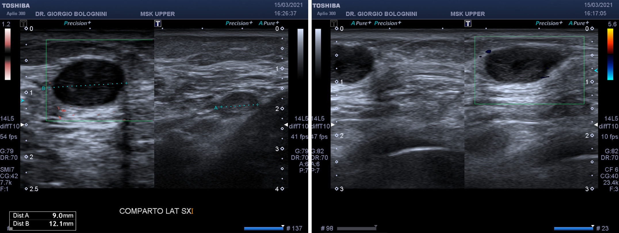
In March 2021 he came to my observation for the regular oncological follow-up; first of all, I note the "persistence immediately posterior to the femoral condyle of an oval hypoechoic nodule with a small anechoic central core of about 12.5x6.8mm, with fine internal trabeculae. Absence of significant internal vascular signals. Immediately more deeply in the perhypothesis, two similar oval nodular formations are visible, this time more isoechoic with a hyperechoic central hilum, therefore more compatible with reactive lymph nodes, 9 and 8mm in long axis, about 12mm distant from the first formation described". I immediately recommend the execution of ultrasound with CEUS contrast for suspicion of recurrence of synovial sarcoma.

The CEUS is performed at the S.D. Diagnostic and Interventional Ultrasound in Cisanello transplants (director Dr. A. Campatelli) with the following result: "The left popliteal palpable swelling is shown ultrasoundally as a hypoechoic oval formation of the subcutaneous plane of 14x8mm, with modest peripheral vascularization only, and at CEUS shows hypoenhancement in all phases; this behavior is not typical of reactive lymph nodes, for which excisional biopsy is recommended". The result of the biopsy is unfortunately a recurrence of synovial sarcoma (sinovial sarcoma).
Histologically, the operative piece appears to be made up of solid whitish material, with myxoid areas. An important radicalization intervention is then carried out in July 2021, again at the Oncological and Reconstructive Orthopedics Unit of Careggi (director Prof. Domenico Andrea Campanacci).
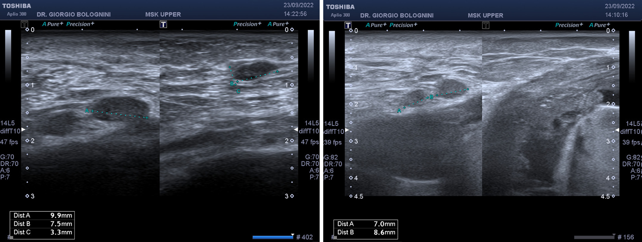
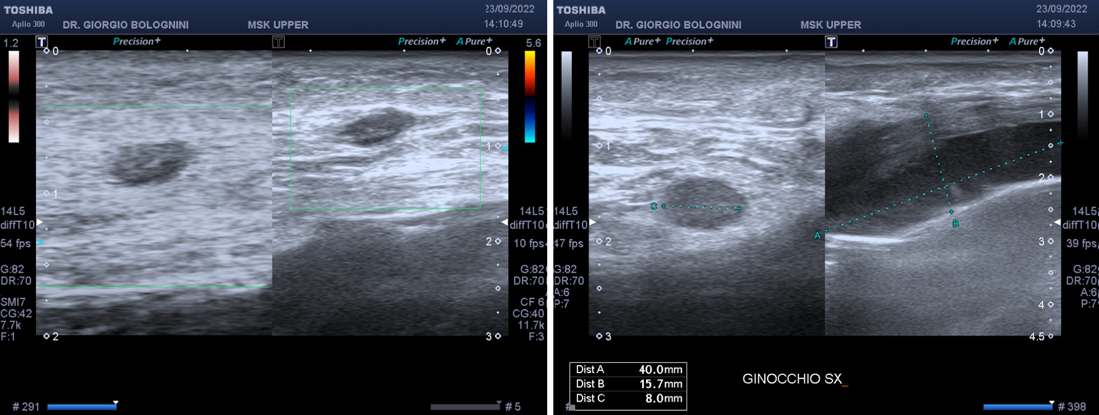
In September 2022 he returned to my ultrasound clinic for an extra-hospital instrumental follow-up, with the following result: "The ultrasound study of the upper posteromedial compartment of the left knee shows the presence of a significantly hypoechoic oval nodule affecting the supramuscular soft tissues, of about 7.5 x3.3mm, of non-unique interpretation. The two similar oval nodular formations more isoechoic with hyperechoic central hilum, therefore compatible with reactive lymph nodes, of 9 and 8 mm in long axis are more deeply stable in perhypothesis. The lateral periprosthetic effusion and the post-scar reworking of the hypodermic and muscular tissue in the popliteal cavity and on the posterolateral side of the ipsilateral leg are still significant. Synovial hippsexation of about 15mm with intra-articular villous femorotibial morphology. The pericicatricial tissues are free from disease recurrence"

The patient was immediately subjected to a new excisional biopsy at the Careggi Oncological Orthopedics which showed the presence of a second recurrence of synovial sarcoma. The patient is currently waiting for a new surgical radicalization.