- Dott. Giorgio Bolognini
- Clinical Cases
- Apr 2023
CASE 12 - Omental cake - Peritoneal carcinomatosis
Peritoneal carcinomatosis
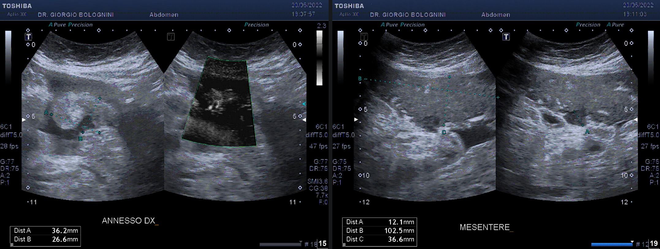
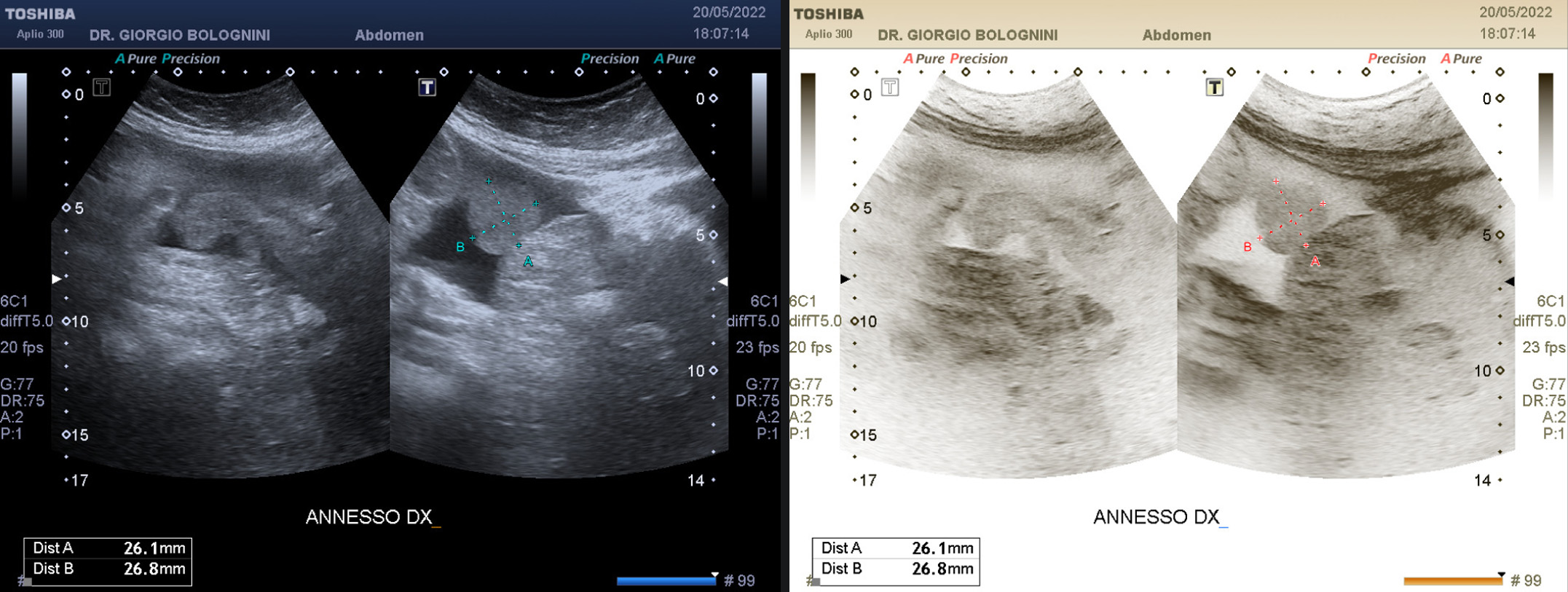
62-year-old woman, two pregnancies with natural delivery, operated on in the past for uterine fibroids; does not take chronic drugs; has been going to my clinic for a general malaise associated with a globular abdomen for several months despite a strict diet, followed monthly by a nutritionist; the woman shows recent haematological evidence of alteration of tumor markers, in particular of CA 125 (CEA 8.10, CA 15.3 215.10, CA 125 1215, CA 19.9 0.20). I then perform an abdominal ultrasound, identifying in the context of a modest ascitic effusion involving all the abdominal recesses a right adnexal mass magnified by the effusion of about 36x26mm, equipped with its own vascularization of an aberrant type, and solid plastron of the meso-hypogastric peritoneum with a maximum thickness of 36mm, of the type omental cake, with hypoechoic peripheral solid nodulation of about 12mm. Uterus basically within limits. In this context, some small hypoechoic nodules of the left hepatic lobe were also highlighted, the largest of about 4 mm at the margin of the III L and 6 mm at the level of the IV L, suggestive of secondary symptoms. Voluminous pathological lymph node of about 18mm in the hepatic hilum.


The appearance of peritoneal carcinomatosis resembled an extensive and thick flow of neoplastic tissue, with an inhomogeneous density, interposed between the abdominal wall and intestinal loops. Maximum tissue thickness was approximately 3.6cm, with a cranial-caudal extension of at least 12cm; from the images it is possible to clearly highlight the origin of the tissue from the parietal peritoneum, with extension in depth towards the loops.
The woman urgently performed an abdominal CT with contrast medium which confirmed the picture of gross peritoneal carcinomatosis (diffuse thickening of the peritoneal leaflets with a scirrhous appearance) with liver metastases and right adnexal mass as the presumable origin of the disease. The woman went to the IEO in Milan where she underwent cisplatin-based cytoreduction with intraperitoneal chemohyperthermia (HIPEC) in anticipation of a peritonectomy. Unfortunately, the patient died of cardiac complications related to the treatment