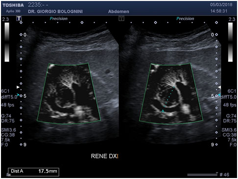
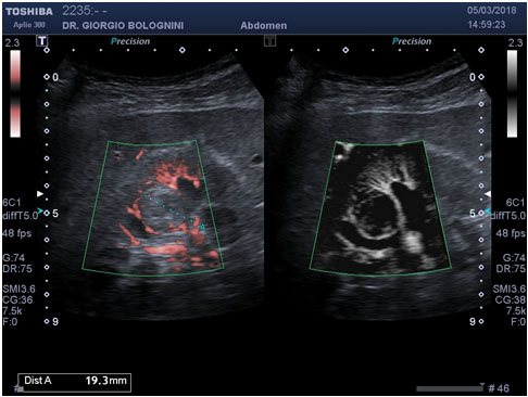
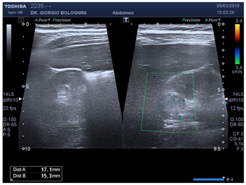
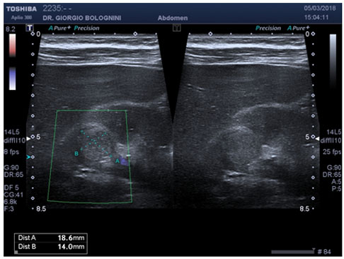
A 55-year-old man in excellent health, BMI 22, came for an abdominal check due to prostate symptoms (dysuria and perineal discomfort) that had been going on for about a month, with laboratory tests (PSA tot 2.50 µg/L, ESR 2, creatinine 0.80 mg/dl, eGFR 88 ml/min) and normal urine (proteins, blood and urinary nitrites absent). The patient had no pathological weight loss or lack of appetite, was apyretic and had no significant changes in mood. The abdominal ultrasound study carried out with extreme accuracy, thanks also to the patient being slim, identified at the level of the right anterior mesorenal region, inside a renal column thus close to the breast, a roundish focal formation with nuanced margins, slightly iso-hyperechogenic, roughly 17x14 mm. .


The formation, poorly identifiable with simple coronal and transverse scans, seemed to affect the lower marginal deep region of a renal column, between two sections, inducing a sort of compression on them; the microvascular study showed an intense peripheral signal and interruption of the normal substantially rayed vascular design of the inter-lobes; internal vascular signals were scarce if not null. There was no involvement of the right renal vein and the vena cava was physiologically collapsible during respiratory testing. Given the anterior mesorenal position and by virtue of the patient's excellent BMI, I also carried out an approach with a high-frequency linear probe; in this case, more modestly hypoechoic internal areas seemed to be appreciated, in any case of a non-cystic fluid type.


I very cautiously diagnose a small solid renal lesion interrupting the normal parenchymal echo-structure of uncertain significance, worthy of study of level II with targeted MRI. The close proximity to the renal sinus also posed differential diagnosis problems with possible urothelial carcinoma. The patient carried out two other eco-color-doppler checks with ultrasounds not equipped with microvascular Doppler, which did not detect significant lesions of the right kidney. However, we proceeded with an MRI investigation that instead confirmed the presence of the lesion, defining it as a focal lesion with a complicated cyst type with internal solid particles. The patient was then operated the following month with enucleation of the lesion. The pathological finding confirmed the malignant nature of the lesion: small intracystic clear cell carcinoma (Grading pT1N0M0), unfortunately with positive enucleation margins. The patient follows a quarterly ultrasound follow-up at my laboratory.