A 31-year-old man, positive familiarity with polycystic kidney disease (mother and sister); no other significant elements to be reported both in family history and in remote and nearby pathological history. BMI 20. He is sent directly by his doctor to my ultrasound laboratory for non-specific disorders of the general malaise type: no fever, blood test results within the norm, no pathological weight loss, regular appetite, no significant changes in the intestine tract; no changes in mood. Regular urination. The abdominal ultrasound study conducted both with a convex probe and with a linear probe thanks to the slim physical constitution of the patient allow me to identify a right renal focal formation, isoechogenic, of about 14 mm, in the upper central mesorenal position, leaning against the breast (at the level of the lower margin of the renal column). The echo-structure shows homogeneity in the distribution of echoes. The SMI microvascular study detects only peripheral signals, with interruption of the rayed course of the interlobular arteries. The linear probe approach shows, at the posterior margin of the lesion, a slight hypoechogenicity of a substantially oval shape, of just 1.5 mm.
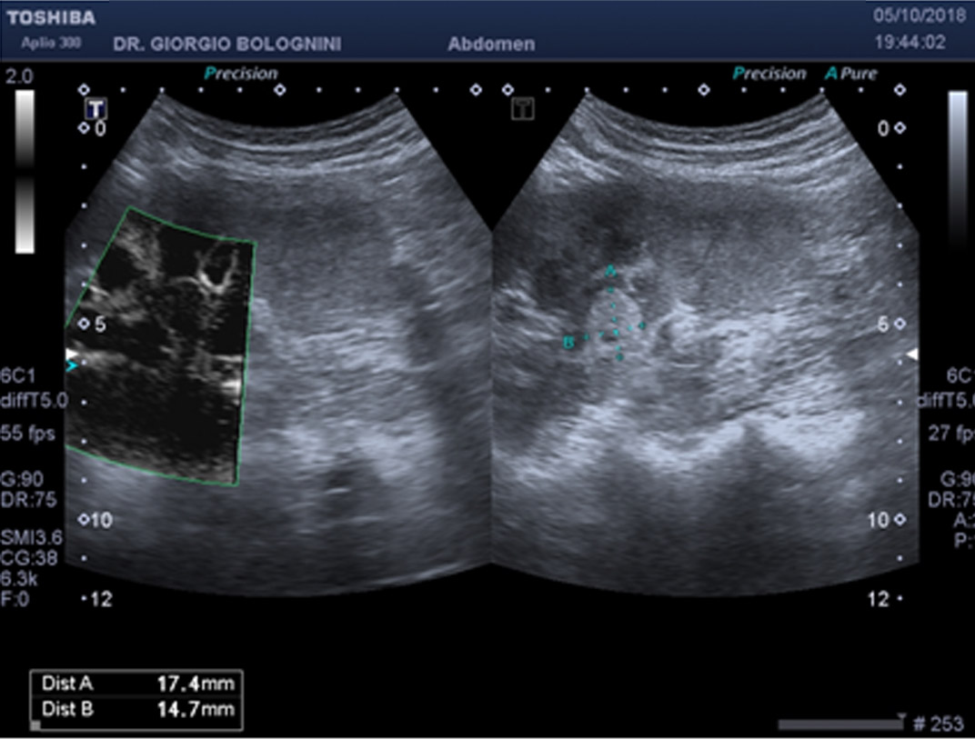
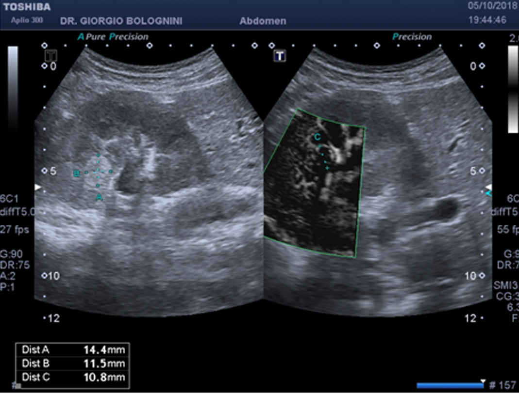
Given his young age, I decide to take him directly to the level II ultrasound laboratory, which essentially confirms the diagnosis of an angiomyolipoma-type focal hyperechoic lesion of the right mesorenal kidney, to be further investigated with a targeted MRI method. Recent MRI demonstrates the complex cystic nature of the lesion (Bosnian III cyst with internal solid tokens). The patient comes to the urological department of Careggi hospital, which essentially postpones any surgical excision until the next MRI check, to be carried out within six months.
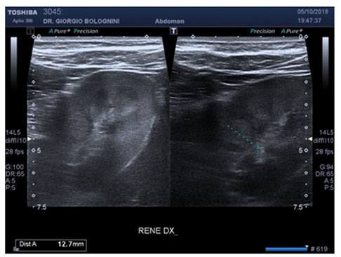
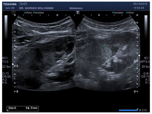
The patient postpones the checks for about 18 months, and then returns to my ultrasound clinic; the examination conducted in particular for the morphological study of the complex angiomyolipoma of the right kidney showed a significant growth of the known lesion with a low adipose component, which went from 15x14mm to approximately 24x23mm, with an evident mass effect on the surrounding arc-shaped arterioles and the presence of internal vascular signals under study with SMI microvascular Doppler.
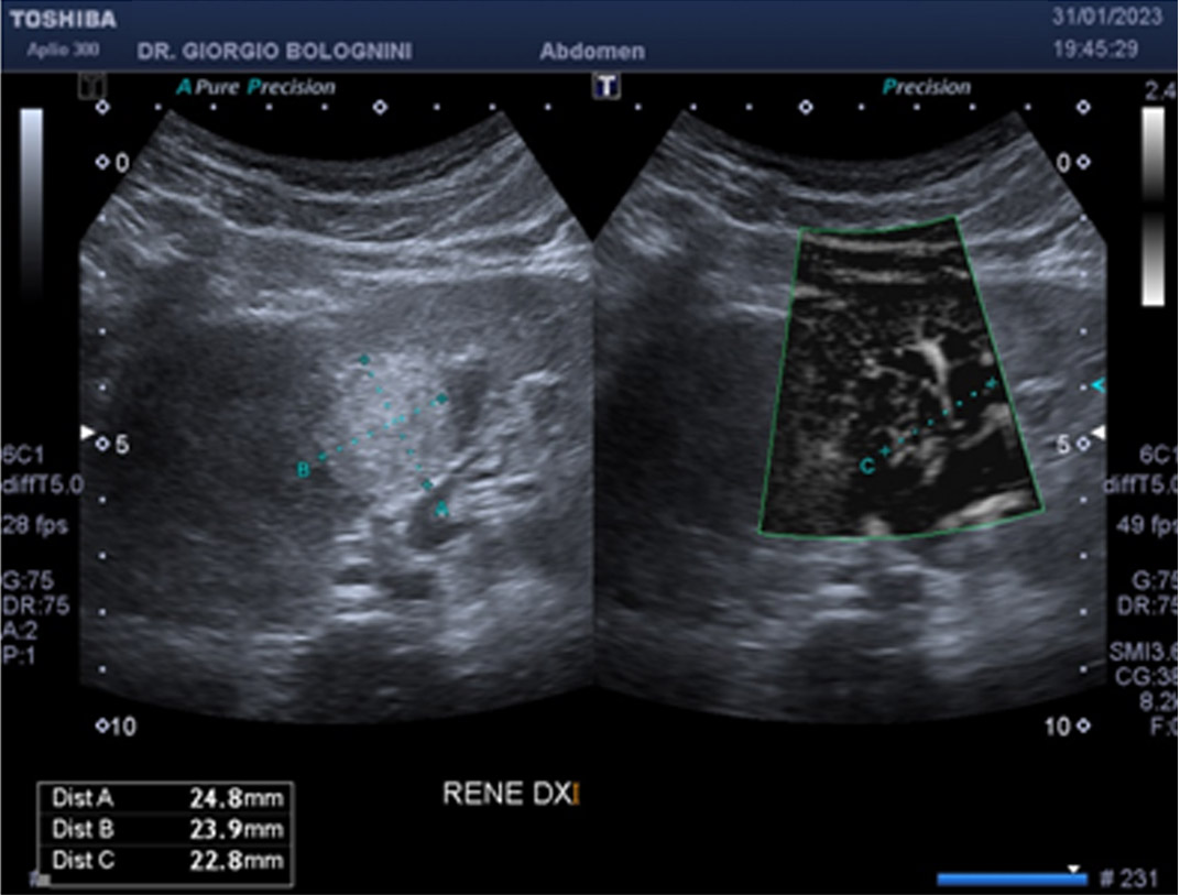
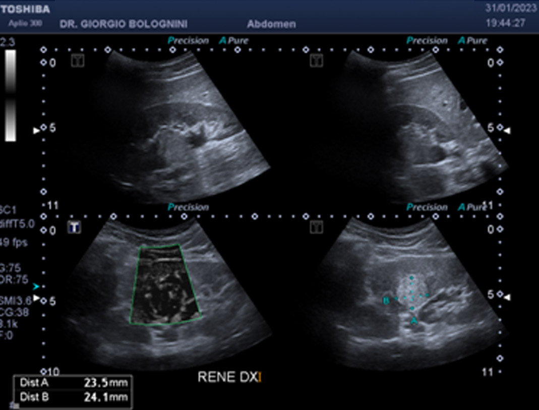
I therefore invite the patient to carry out a level II diagnostic test with contrast by contacting hospital urology again. After two weeks, the patient then performed an abdominal CT + contrast medium which confirmed the presence of a "nodular lesion on the right mesorenal bone, with irregular margins, with marked post-contrast enhancement, of 24x22mm in maximum diameter, protruding at the level of the pyelic sinus and compressing and displacing the medium calyces, therefore compatible with dyskaryokinetic lesion. No signs of thrombosis at the level of the ipsilateral renal vein". The patient is then operated on after about a month by Professor Andrea Minervini, Director of the complex departmental unit of Minimally Invasive Robotic and Andrological Oncological Urology at A.O.U. Careggi, which performs a transrobotic enucleation with lumbar access with preservation of the organ. The subsequent histology result is as follows: Clear cell renal carcinoma, nucleolar grade 3 (WHO 2022), absent necrosis, perilesional adipose tissue and surgical margins intact, pT1a staging.
The patient is therefore undergoing post-surgical oncological screening at my ultrasound clinic every six months.